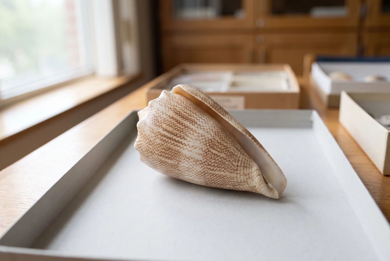
Two peptides can have the same amino-acid sequence and intact mass while their disulfide bonds connect different cysteines. Studies of cone-snail peptides show that these alternative pairings can change laboratory activity and even overturn an assumed structure. Identifying the bridges requires evidence beyond a mass match.[1][3]
Buy 1, Get 1 20% Off
Pick any two research compounds. Take 20% off the lower priced one.
Applied automatically at checkout, no code needed
Claim my 20% off →In every pair, the lower priced item is 20% off after other discounts; accessories excluded. For research use only.
Science explainer. The conotoxins discussed here are experimental probes studied in laboratory systems, not treatment recommendations.
The same bridge count can connect different partners
A disulfide bond joins the sulfur atoms of two cysteine residues, the cysteine building blocks within a peptide. When several cysteines are present, the same chain can support more than one pairing pattern. Chemists call these disulfide-connectivity isomers: the sequence is unchanged, but the bridges join different positions.[1][2]
Imagine four cysteines numbered in the order they appear along a chain. They can form two bridges by pairing the first with the third and the second with the fourth. Pairing the first with the fourth and the second with the third also uses all four cysteines and makes two bridges. A third pattern pairs neighboring cysteines. The conventional names are globular, ribbon and beaded, respectively.[2]
Changing partners in this way changes the connectivity without changing which atoms the molecule contains. Fully oxidized forms with the same bridge count can therefore share an intact mass. Their shapes and interactions can still differ because different parts of the chain are held together.[1][2]
This is a different question from whether a peptide is cyclic or linear. The cyclic-peptide explainer examines what closing a ring can do to stability and cell entry. Here, the molecules can already contain the same number of bridges; the unresolved detail is which cysteines those bridges connect.[1][3]
Two chromatography peaks with the same mass
In a 2012 study, researchers made the cone-snail peptide KIIIA and separated its laboratory folding products by high-performance liquid chromatography, or HPLC. Two distinct peaks appeared. Both fractions had the same measured intact mass, consistent with the same sequence and three disulfide bonds.[3]
Chromatography had detected a difference that the intact mass did not identify. The more abundant product emerged from the column before the less abundant one, showing that the two forms behaved differently during separation. That observation alone did not reveal their bridge pattern.[3]
The team then examined fragments produced from the disulfide-linked material. Those fragments constrained which cysteines could be connected. For the major KIIIA product, further fragmentation established the linkage pattern directly. Assignment of the minor product also used the distinct retention time and the observation that reducing both forms yielded products that co-eluted.[3]
Reduction opens disulfide bonds. Once those bridges were removed, the two fractions no longer showed the same chromatographic distinction. Combined with the fragment evidence, that helped the researchers distinguish a change in bridge pairing from a different underlying peptide chain.[3]
A plausible structure can still contain an assumed bond
KIIIA also exposed a limitation of interpreting one structural method in isolation. An earlier nuclear magnetic resonance, or NMR, structure had used a disulfide pattern inferred from related peptides. The bond-mapping study showed that the major laboratory folding product used a different pattern.[3]
When the researchers recalculated the structure, both the earlier assumed pattern and the experimentally supported pattern could fit the existing NMR data. The overall structure changed little, and a helical region containing important channel-blocking residues remained. A convincing-looking fold was therefore not sufficient to settle the bridges.[3]
That is why a structural assignment needs to distinguish measured constraints from assumptions used to build a model. NMR can contribute detailed information about shape, but an underconstrained set of observations may allow more than one connectivity. Direct chemical evidence can resolve an ambiguity that the model's appearance cannot.[3]
Native venom adds another complication
Multiple forms are not confined to a synthetic reaction. A 2023 study of PnID, a peptide from Conus pennaceus, found three closely eluting signals at the expected mass-to-charge value in a venom-duct extract. Treating the material with a reducing agent brought the signals together into one corresponding peak.[2]
The researchers then prepared and characterized synthetic forms. They assigned globular and ribbon connectivity to the two main products of a nonselective folding experiment, using chromatographic comparisons and chemical bond analysis. The native extract had shown three peaks, while that laboratory preparation produced two prominent forms.[2]
The third native peak remained uncertain. The paper discusses a beaded arrangement, a dimer and other structural possibilities, without conclusively assigning every native peak. Native venom can contain more structural variety than a single expected-mass signal suggests, but those peaks still need individual identification.[2]
Pairing changes activity, but there is no universal winning shape
For TxIA, the globular form blocked a rat nicotinic acetylcholine receptor more strongly than the ribbon form, while the beaded form showed little activity in that assay. The receptors were expressed in Xenopus frog egg cells so researchers could measure electrical currents. This was a controlled receptor experiment, not an observation of benefits in a person.[1]
PnID gave a different ordering. Its ribbon form was the most active at inhibiting norepinephrine uptake in rat-brain synaptosomes, preparations containing nerve endings. The globular and beaded forms were much less effective in that test. The target and the peptide differed from TxIA, so the topology names alone could not predict the result.[2]
KIIIA complicates an all-or-nothing interpretation too: both major and minor synthetic isomers blocked the rat sodium-channel subtype NaV1.2 in frog egg cells, although their measured strengths differed. An alternative pairing did not automatically erase activity.[3]
Together, these experiments show why mass, connectivity and function are separate questions. An activity assay can reveal a difference worth investigating, but activity alone cannot identify the bridge pattern. These laboratory results also cannot establish human safety or therapeutic benefit.[1][2][3]
Which measurement answers the structural question?
- Intact mass tests whether the detected molecular mass matches an expectation. It cannot uniquely distinguish disulfide pairings that preserve composition and bridge count.[1][3]
- Chromatography can separate some isomers, as it did for KIIIA. Closely eluting TxIA forms show why separation depends on the molecule and the analytical method.[1][3]
- Fragment analysis and selective disulfide analysis can locate connections. Their conclusions depend on which bonds and fragments were actually resolved.[2][3]
- NMR adds evidence about three-dimensional structure. The KIIIA example shows why researchers must check whether an assumed connectivity is independently constrained.[3]
A mass match is the beginning of an assignment
These papers used complementary measurements because no single readout answered every question. TxIA's native peptide was compared with characterized synthetic forms; PnID combined chemical analysis with NMR; KIIIA required fragment interpretation and chromatographic evidence to revisit a prior structural assignment.[1][2][3]
The practical scientific consequence is an interpretable activity comparison. Before attributing a change in receptor behavior to a sequence, researchers need to know which disulfide-connected form was tested. Matching the mass keeps one part of the identity consistent. Establishing the bridges supplies the structural detail needed to explain why the same sequence can behave differently.[1][3]
Frequently asked questions
Can chromatography distinguish disulfide isomers?
Sometimes. The KIIIA study separated two isomers with the same intact mass. TxIA forms could have similar retention times and required careful comparisons. A separate peak is evidence of a difference, but it does not by itself name the disulfide connections.
Is the most abundant laboratory folding product necessarily the native form?
No. The KIIIA researchers identified the major synthetic product but left open whether its connectivity matched the peptide in cone-snail venom. Biological folding can involve precursor sequences, folding enzymes and molecular chaperones that are absent from a simple laboratory preparation.
Sources and further reading
Related reading
Buy 1, Get 1 20% Off
Pick any two research compounds. Take 20% off the lower priced one.
Applied automatically at checkout, no code needed
Claim my 20% off →In every pair, the lower priced item is 20% off after other discounts; accessories excluded. For research use only.