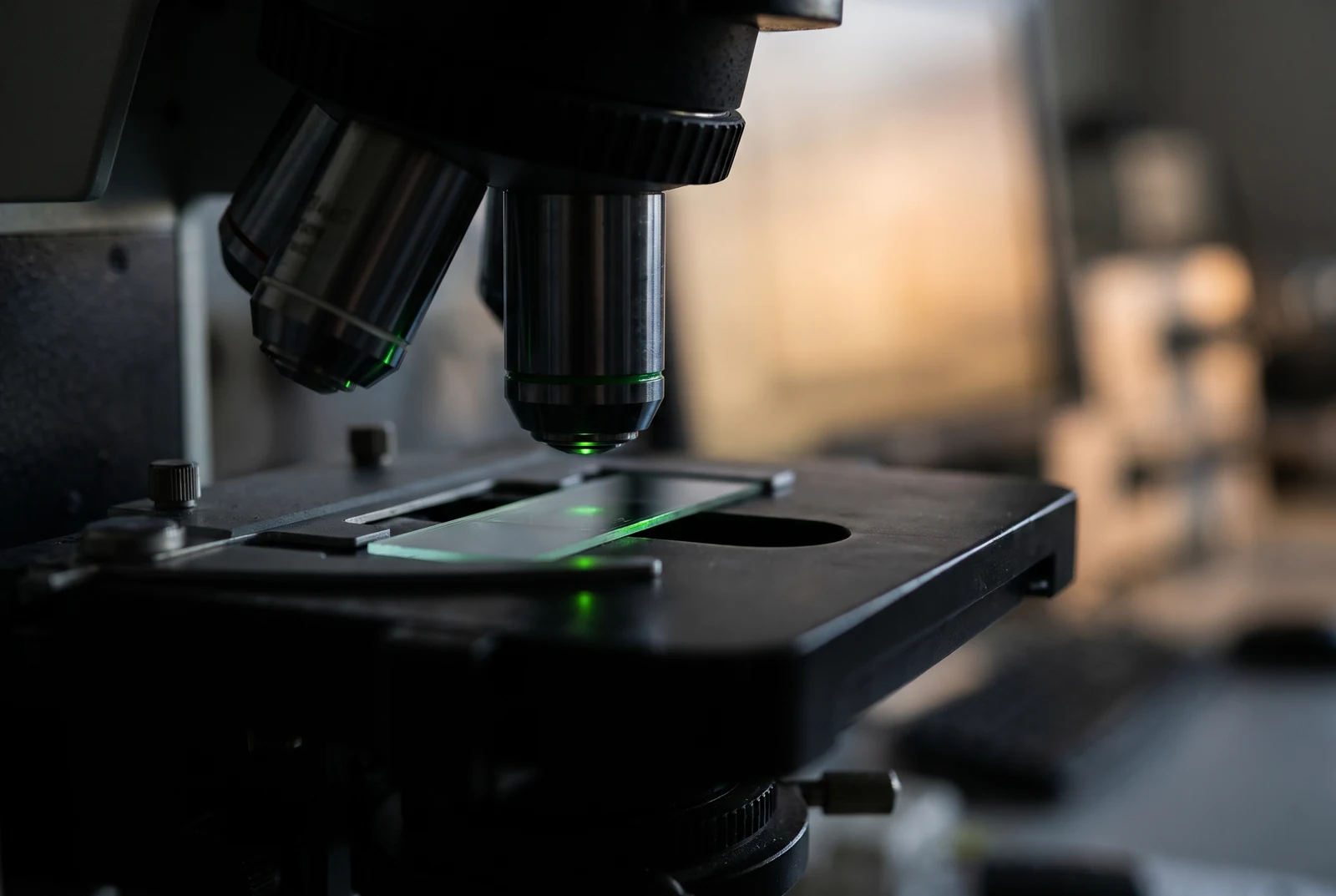
A fluorescent label can change how a peptide behaves. Experiments have found altered membrane interactions, different molecular shapes and a reversal between cell and animal imaging results after changing the attached dye. The signal locates fluorescent material; whether that material represents the original peptide is a separate experimental question.[1][2][3]
Buy 1, Get 1 20% Off
Pick any two research compounds. Take 20% off the lower priced one.
Applied automatically at checkout, no code needed
Claim my 20% off →In every pair, the lower priced item is 20% off after other discounts; accessories excluded. For research use only.
IQON Labs' research use only catalog is separate from these experiments. The studies do not evaluate the catalog.
The label becomes part of the molecule
Attaching a dye makes a peptide easier to see, but also adds a chemical group with its own charge, size and attraction to water or lipid. That creates two questions for an imaging experiment: where the labeled material goes, and whether the label helped send it there. Work on penetratin, a peptide studied for cellular delivery, shows why those questions cannot be separated by choosing a brighter microscope setting.[1]
In a 2018 Scientific Reports study, researchers compared unlabeled penetratin with versions carrying six different fluorescent labels. They examined interactions with artificial lipid membranes using several methods, including measurements of leakage, binding heat and membrane structure. The comparison kept the underlying peptide sequence constant while changing the attached reporter.[1]
A membrane experiment changes with the dye
Unlabeled penetratin mainly associated with the membrane surface and produced little disruption of its hydrophobic interior. The more hydrophobic rhodamine B and pyrenebutyric acid conjugates penetrated further into the model membranes and caused greater disturbance. Structural measurements supported changes such as membrane thinning and loss of lipid material.[1]
The team also trapped a fluorescent substance called calcein inside lipid vesicles. Its escape supplied a readout of membrane leakage. The rhodamine B and pyrenebutyric acid conjugates released much more calcein than the unlabeled peptide under the conditions tested. That result concerned artificial membranes, not proof of improved delivery into living cells.[1]
Some labels created a second difficulty: they interfered with the measurement itself. The carboxyfluorescein conjugate emitted at the same wavelength as calcein, while the TAMRA conjugate could absorb some of calcein's emitted light. The researchers could not obtain reliable leakage results for those two conjugates. A dye can change the specimen and also obstruct the test used to measure that change.[1]
The reporter can hold a peptide in a different shape
A 2017 PLOS ONE study examined a different system: a short, flexible fragment of the ribonuclease S peptide. The investigators attached Atto655 dye at one end and added tryptophan, which quenches fluorescence on contact, at the other. Fluctuations in light were intended to reveal how the ends moved toward and away from each other.[2]
Molecular simulations suggested that aromatic rings in the dye and tryptophan could stack together, stabilizing contact between the ends. In that model, the labeled construct explored different shapes and rearranged more slowly than the unmodified peptide. Measurements of circular dichroism, which probe structural populations, also supported a change in the mixture of structures.[2]
The result applies to this dye and quencher construct. It does not isolate the effect of the dye from that of the added tryptophan, and the authors acknowledged limits to the conformations their simulations could sample. Even so, it exposes a specific interpretive trap: local dye contact can be measured accurately while giving an incomplete account of the motion of the whole chain.[2]
Strong cell fluorescence need not predict an animal image
In a 2021 Cancers paper, Takakura and colleagues coupled the same experimental peptide to cyanine dyes or their sulfonated counterparts. Sulfonation changed the conjugates' physicochemical properties. In cultured human pancreatic cancer cells, the less hydrophilic Cy3 and Cy5 conjugates produced stronger cellular fluorescence than the corresponding sulfonated versions.[3]
The ordering changed in mice carrying tumors derived from the same pancreatic cancer cell line. The Cy5 conjugate gave little tumor fluorescence, whereas the sulfoCy5 conjugate produced a clearer tumor signal. The cell culture result had not identified a universally better label. Distribution through an animal introduced a different set of conditions.[3]
In a separate experiment, the researchers incubated the Cy5 and sulfoCy5 conjugates in plasma collected from mice. Chromatography, which separates a mixture's components, showed rapid breakdown of both conjugates. On that basis, the authors suggested that degradation products accounted for much of the fluorescence in the animal images.[3]
The paper was corrected in 2022 to remove a peptide name and sequence published without the inventors' agreement. It now identifies the material as peptide A. The correction states that the scientific conclusions were unchanged, but the missing sequence limits how fully a reader can reconstruct the molecule from the article.[4]
Connect the image to a chemical measurement
These experiments point to different checks for different claims. Comparing a labeled peptide with its unlabeled counterpart in a functional assay asks whether the reporter preserves the behavior of interest. Comparing several labels can reveal dependence on dye chemistry. Separating and identifying intact conjugate and fragments addresses whether the light still belongs to the intended molecule.[1][2][3]
None of those checks alone settles where material sits inside a cell. That requires evidence about cellular compartments. The earlier question is whether the chemical object being tracked still represents the peptide the experiment set out to study.[1][3]
For the clearer sulfoCy5 tumor image, the unresolved question is how much light came from intact conjugate and how much from breakdown products. Chemical analysis of the fluorescent material in the tumor would help distinguish delivery of the labeled peptide from accumulation of its fragments.[3]
Sources and further reading
- Fluorophore labeling of a cell-penetrating peptide significantly alters the mode and degree of biomembrane interaction. DOI: 10.1038/s41598-018-24154-z
- Covalent dye attachment influences the dynamics and conformational properties of flexible peptides. DOI: 10.1371/journal.pone.0177139
- In Vitro and In Vivo Cell Uptake of a Cell-Penetrating Peptide Conjugated with Fluorescent Dyes Having Different Chemical Properties. DOI: 10.3390/cancers13092245
- Correction: Takakura et al. In Vitro and In Vivo Cell Uptake of a Cell-Penetrating Peptide Conjugated with Fluorescent Dyes Having Different Chemical Properties. Cancers 2021, 13 , 2245. DOI: 10.3390/cancers14081880
Related reading
Buy 1, Get 1 20% Off
Pick any two research compounds. Take 20% off the lower priced one.
Applied automatically at checkout, no code needed
Claim my 20% off →In every pair, the lower priced item is 20% off after other discounts; accessories excluded. For research use only.